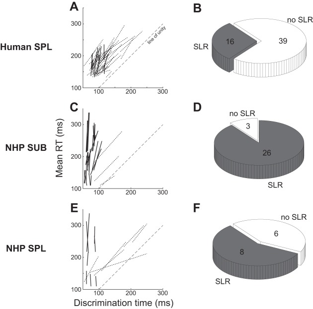
Fig. 4.
SLR prevalence on SPL in humans (A and B), on the suboccipital muscles in NHPs (C and D), and on SPL in NHPs (E and F). A, C, and E show the lines connecting the points for the short and long RT groups for all subjects, with solid or dashed lines conveying instances where the SLR was or was not detected, respectively. The pie charts in B, D, and F depict the prevalence in the SLR in the various samples, and provide the no. of observations as well.

