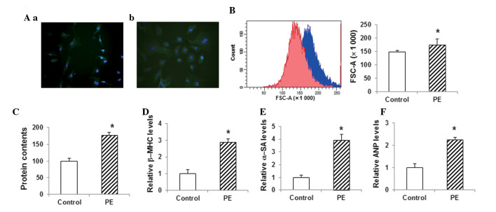
Figure 2.
Construction and identification of the in vitro model of myocardial hypertrophy. (A) Primary myocardial cells from 3-day-old rats were cultured (a) with or (b) without 100 µM PE (magnification, x400). Cells were stained with α-SA antibody (green) and DAPI (blue). (B) Sizes of cardiac myocytes in the control group (red) and the PE-treated group (blue) were detected using flow cytometry. (C) Total cellular protein levels were determined using the bicinchoninic acid assay. (D-F) The mRNA expression levels of the hypertrophy-related genes (D) β-MHC, (E) α-SA and (F) ANP in myocardial cells were detected via the quantitative polymerase chain reaction. Compared with the control group, *P<0.05. PE, phenylephrine; β-MHC, β-myosin heavy chain; α-SA, α-sarcomeric actinin; ANP, atrial natriuretic peptide; FSC, forward scatter.

