Figure 4.
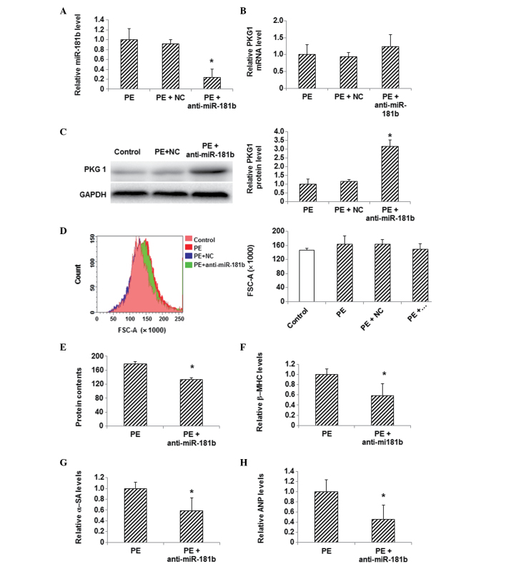
Effects of miR-181b inhibition on PKG 1 expression and hypertrophy in PE-treated myocardial cells. (A) The expression level of miR-181b was detected via qPCR in myocardial cells following the transfection of miR-181b inhibitor. (B) mRNA and (C) protein expression levels of PKG 1 were detected in myocardial cells using qPCR and western blotting, respectively, following miR-181b inhibitor transfection. (D) Sizes of cardiac myocytes in the control (pink), PE (red), PE + NC (green) and PE + anti-miR-181b (transfected with miR-181b inhibitor) (blue) groups were detected with flow cytometry. (E) Total cellular protein levels were determined using the bicinchoninic acid assay. (F-H) The mRNA expression levels of the hypertrophy-related genes (F) β-MHC, (G) α-SA and (H) ANP in myocardial cells following transfection were detected using qPCR. Compared with the PE-treated group, *P<0.05. miR, microRNA; PE, phenylephrine; NC, random sequence; PKG 1, cGMP-dependent protein kinase type I; β-MHC, β-myosin heavy chain; α-SA, α-sarcomeric actinin; ANP, atrial natriuretic peptide; FSC, forward scatter; qPCR, quantitative polymerase chain reaction.

