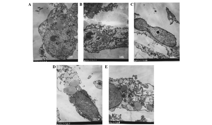
Figure 4.
Transmission electron microscopy of ultrastructural changes in the A431 cells treated with varying concentrations of EPS-A. (A) The control cells were treated with phosphate-buffered saline for 48 h. (B) Nuclear fragmentation, chromosome condensation, cell shrinkage and loss of membrane integrity were observed in the A431 cells treated with 1 mg/ml EPS-A for 48 h; (C) severe chromosome condensation and cell shrinkage were observed in the A431 cells treated with 2 mg/ml EPS-A for 48 h; (D) expansion of the endoplasmic reticulum was observed in the A431 cells treated with 2 mg/ml EPS-A for 48 h, and (E) apoptotic bodies were observed in the A431 cells treated with 3 mg/ml EPS-A for 48 h (scale bar=1 micron). EPS-A, extracellular polymeric substances of Aphanizomenon flos-aquae.

