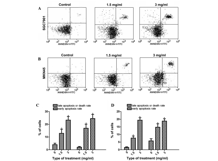
Figure 2.
Flow cytometric analysis of PI-annexin-V to quantify Huaier-induced apoptosis in (A and C) SGC7901 and (B and D) MKN45 cells. Dot plots show the results following the treatment of the SGC7901 and MKN45 cells with Huaier at concentrations of 0, 1.5 and 3 mg/ml for 24 h. The experiment was performed in triplicate, and the data are expressed as the mean ± standard deviation of the three separate experiments. *P<0.05, compared with the control. PI, propidium iodide; FITC, fluorescein isothiocyanate.

