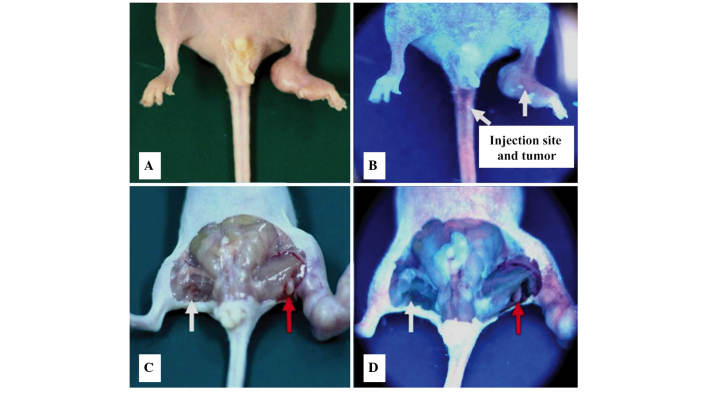
Figure 4.
Tracking of deuteporfin to the tumor sites of the lymph node metastatic model of a pancreatic cancer cell line under irradiation of a Wood's lamp. Mice 1 h after intravenous administration of deuteporfin (A) without and (B) with the Wood's lamp. Excised mice 1 h after intravenous administration of deuteporfin (C) without and (D) with the Wood's lamp. White arrows indicate the normal popliteal fossa lymph nodes and the red arrows indicate the metastatic lymph nodes. Blue fluorescence indicates the effect of the Wood's Lamp.

