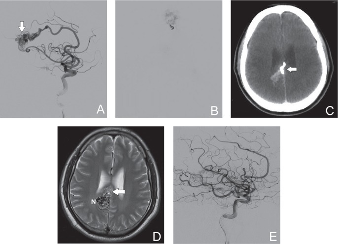
Fig. 1.
A 50-year-old male with a ruptured AVM (Spetzler-Martin grade III). A: DSA (lateral view, right internal carotid angiography) shows the nidus (arrow) and main feeding artery from the right anterior cerebral artery. B: Injection of Eudragit-E is clearly visible with fluoroscopy. C: Postoperative CT image (axial view) shows the cast of Eudragit-E (arrow) with minimal artifacts. D: Postoperative T2-weighted MRI (axial view) reveals the Eudragit-E cast (arrow) and residual nidus (N). E: Postoperative DSA image (lateral view) after two sessions of endovascular embolization and gamma knife radiosurgery shows no residual AVM. AVM: arteriovenous malformation, CT: computed tomography, DSA: digital subtraction angiography, MRI: magnetic resonance imaging.

