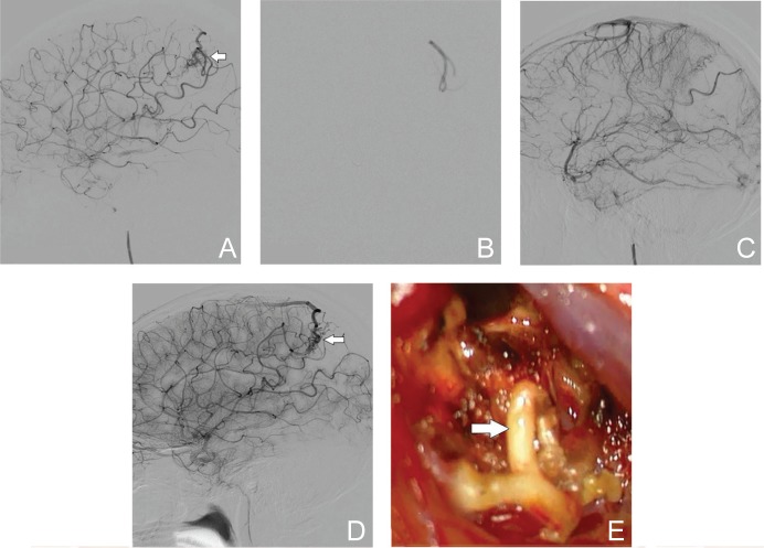
Fig. 2.
A 16-year-old female with an intracerebral hemorrhage from a ruptured AVM (Spetzler-Martin grade II). A: DSA (lateral view, left internal carotid angiography) reveals the nidus (arrow) with feeders from the left angular artery. B: Injection of Eudragit-E is clearly visible with fluoroscopy. C: Postoperative DSA (lateral view) shows no residual AVM. D: Follow-up DSA performed 6 months later reveals the recurrence of AVM (arrow). E: Blood vessels that were embolized with Eudragit-E (arrow) are elastic and whitish in color. AVM: arteriovenous malformation, CT: computed tomography, DSA: digital subtraction angiography.

