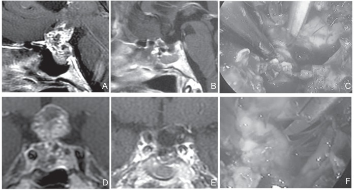
Fig. 1.
A, D: Preoperative sagittal (A) and coronal (D) T1-weighted post Gd MRI. The tumor was of subdiaphragmatic type. The sella turcica was enlarged. The tumor extends in the coaxial direction, similar to the approach. The sphenoid sinus was also enlarged. No lateral extension not beyond the internal carotid artery wasobserved. This tumor effectively underwent EETSA. C, F: Perioperative photograph. The operation was easy because fenestration from the planum sphenoidale to the sella turcica could be secured. Sharp dissection of tumor from the undersurface of the optic nerve was performed within the visual field of endoscopy. (C) Surgical field through endoscopy during resection was completed (F). B, E: Postoperative sagittal (B) and coronal (E) T1-weighted post Gd MRI. No residual tumor wasobserved. In the case of adequate enlargement of the sella turcica, even when the prechiasmatic space was small, enlarged sella turcica could be effectively isolated. EETSA: endoscopic extended transsphenoidal approach, MRI: magnetic resonance imaging.

