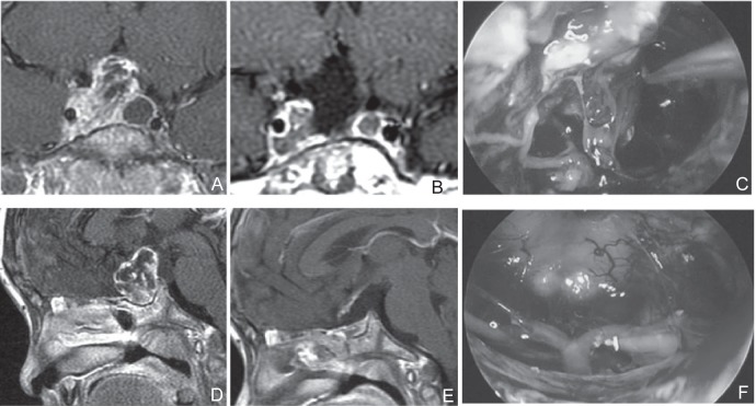
Fig. 2.
A, D: Preoperative sagittal (A) and coronal (D) T1-weighted post Gd MRI. A poorly developed sphenoid sinus, which was suitable for EETSA, although extension to cavernous sinus was suspected. During the UBIHA, it was judged that operation of this area might be difficult and resection was performed using navigation in EETSA. C, F: Perioperative photograph. Resection was performed while confirming the film of tumor (C). Surgical field through endoscopy when resection was completed (F). B, E: Postoperative sagittal (B) and coronal (E) T1-weighted post Gd MRI. Residual tumor was suspected in the cavernous sinus. After surgery, stereotactic radiotherapy was performed. EETSA: endoscopic extended transsphenoidal approach, MRI: magnetic resonance imaging, UBIHA: unilateral basal interhemispheric approach.

