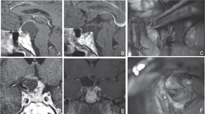
Fig. 3.
A, D: Preoperative sagittal (A) and coronal (D) T1-weighted post Gd MRI of a tumor that occurred in the intrasellar component. The stalk existed in the forward direction. The tumor extended beyond the posterior clinoid process. The prechiasmatic space was wide. Development of the sphenoid sinus was poor but the sellar diaphragm was pushed downward. There was sufficient distance between the optic chiasm and the top surface of the pituitary gland. It was generally judged that EETSA was possible, but UBIHA was chosen in a comprehensive manner because the stalk existed in the forward direction and the tumor extended beyond the posterior clinoid process. C, F: Dissection of a tumor located at the lateral of the internal carotid artery, within the visual field of the UBIHA (C), Dissection of B and A was possible under direct vision in the posterior and inferior areas (F). B, E: Preoperative sagittal (A) and coronal (D) T1-weighted post Gd MRI. Postoperative sagittal (B) and coronal (E) T1-weighted post Gd MRI. Tumor was totally resected. EETSA: endoscopic extended transsphenoidal approach, MRI: magnetic resonance imaging, UBIHA: unilateral basal interhemi spheric approach.

