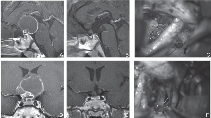
Fig. 4.
A, D: Preoperative sagittal (A) and coronal (D) T1-weighted post Gd MRI of a cystic extension was observed in the anterior cranial base. Although it was predicted that the prechiasmatic cistern was large, UBIHA was chosen because the sphenoid sinus showed poor development, which traversed the distance from the fenestration to the site of operation, and extension to the lateral direction was slightly wide. C, F: Perioperative photograph. Tumor extended beyond the right internal carotid artery (C). Dissection of the oculomotor nerve was necessary (F). B, E: Postoperative sagittal (B) and coronal (E) T1-weighted post Gd MRI, Tumor was totally resected. MRI: magnetic resonance imaging, UBIHA: unilateral basal interhemispheric approach.

