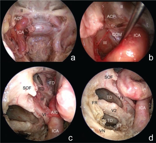
Fig. 2.

Endoscopic anatomy of cavernous sinus triangles. a: The dura mater over the right cavernous sinus is removed and contents of the cavernous sinus are shown. b: The area surrounded by the optic nerve, oculomotor nerve, and ICA is the clinoidal triangle as visualized from the endonasal route. This triangle corresponds with only the anterior area of the clinoidal triangle. An endoscopic view of the clinoid triangle with the anterior clinoid process show the anterior clinoid process located below the optic nerve and lateral to the ICA. Medial displacement of the ICA reveals that the carotid-oculomotor membrane bridging between the oculomotor nerve and ICA attaches to the inferior surface of the anterior clinoid process. c: The clinoid triangle without the anterior clinoid process shows the medial temporal and frontal dura mater through the clinoid triangle. Exposure of the supra- and infra-trochlear triangles is restricted by the abducens nerve, artery of the inferior cavernous sinus, and ICA. d: The anteromedial triangle is defined by the first and second divisions of the trigeminal nerve and a line between the superior orbital fissure and the foramen rotundum. The anterolateral triangle, delimited by the second and third divisions of the trigeminal nerve, is partially recognizable. Because the inferior area of the anterolateral triangle is covered by the sphenoidal bone behind the Vidian nerve, only the superior area of the anterolateral triangle, as delineated by the second division of the trigeminal nerve, proximal segment of the third division of the trigeminal nerve, and the Vidian nerve, is exposed. Inferomedial temporal dura mater is apparent through the entire anteromedial triangle and superior area of the anterolateral triangle.16) ACP: anterior clinoid process, AIC: artery of the inferior cavernous sinus, COM: carotid-oculomotor membrane, FD: frontal dura mater, FR: foramen rotundum, ICA: internal carotid artery, SOF: superior orbital fissure, TD: temporal dura mater, VN: Vidian nerve, V1: first division of the trigeminal nerve, V2: second division of the trigeminal nerve, V3: third division of the trigeminal nerve, II: optic nerve, III: occulomotor nerve, VI: abducens nerve.
