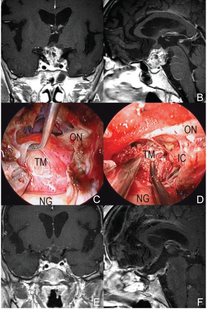
Fig. 3.

A representative case of craniopharyngioma (64-year-old woman with loss of visual acuity). The tumor derived from the pituitary stalk pushed up the chiasm (A, B). In EESB approach, the boundary between the tumor and the optic nerve could be clearly seen and the tumor was removed safely without any injury of optic nerves and surrounding arteries (C, D). Most of tumor was removed on postoperative MRI (E, F). ESSB: endoscopic endonasal skull base, IC: internal carotid, MRI: magnetic resonance imaging, NG: normal pituitary gland, ON: optic nerve, TM: tumor.
