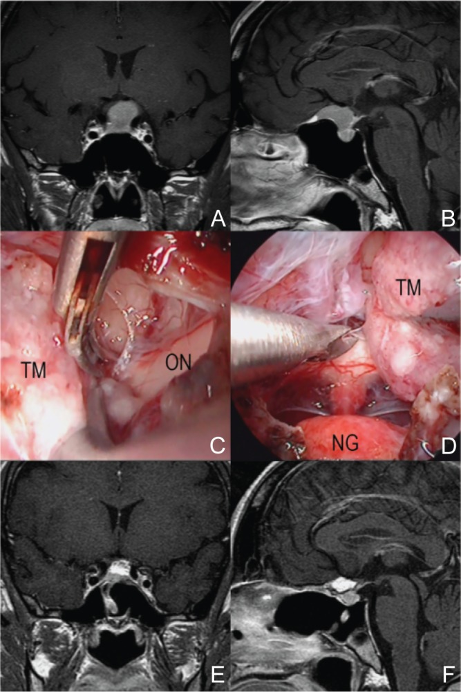
Fig. 4.

A representative case of tuberculum sellae meningioma (37-year-old woman with headache). The tumor attached at the tuberculum and sphenoid planum, compressed the pituitary and the chiasm behind (A, B). During operation, there were no bleeding from tumor and the margin between the tumor and surroundings were clearly seen (C, D). Total removal of tumor was achieved on postoperative MRI (E, F). MRI: magnetic resonance imaging, NG: normal pituitary gland, ON: optic nerve, TM: tumor.
