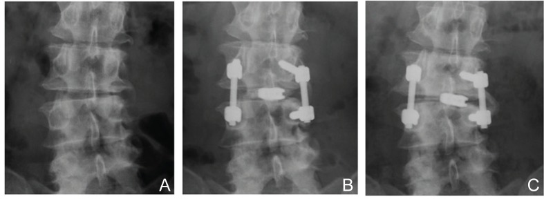
Fig. 6.

A: Preoperative radiograph of a patient with degenerative spondylolisthesis at L3–4 with segmental coronal imbalance. B: Radiograph at 1 week after asymmetrical TLIF revealing correction of lateral disc wedging at the operated site. C: Radiograph at 2 years after operation demonstrating deterioration of segmental and whole lumbar coronal balance with subsidence of the intervertebral spacer. TLIF: transforaminal lumbar interbody fusion.
