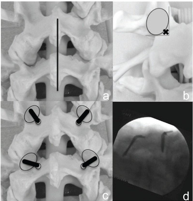
Fig. 3.

Surgical procedure of MIDLF part 1. a: Synthetic model showing midline surgical incision (black line). b: Starting point (x mark) exists at the intersection of medial and caudal aspect of pedicle. c: Synthetic model showing cortical bone trajectory of maker pins. d: Intraoperative lateral image demonstrated the proper position of maker pins. MIDLF: midline lumbar fusion.
