
Fig. 4.
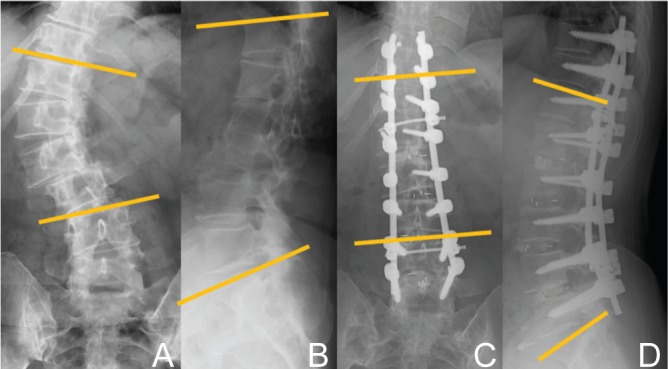
Radiographic studies obtained in a 65-year-old woman who complained of severe low back and radicular pain. A, B: Preoperative radiographs showing 49° of scoliosis (A), 26° of LL (B), 35° of PT, and 59° of PI. Th10–L5 fusion was performed with transforaminal lumbar interbody fusion procedure from L1/2 to L4/L5. C, D: Sixteen months postoperative radiographs showing 3° of scoliosis (C) and 49° of LL angle (D), and 21° of PT. The difference between PI and LL was improved from 33 to 10. LL: lumbar lordosis, PI: pelvic incidence, PT: pelvic tilt.