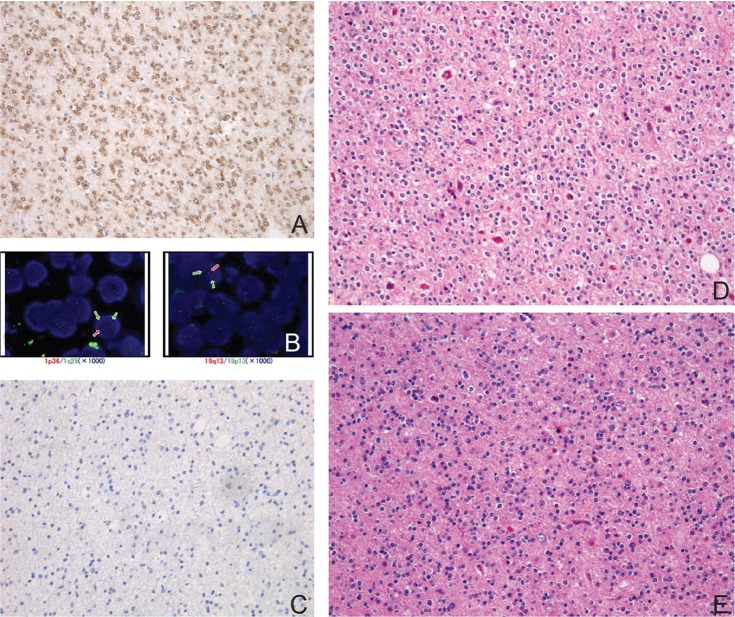
Fig. 2.
Histology of classic oligodendroglioma with double-positive genetic signature. A: Immunohisto-chemistry with isocitrate dehydrogenase (IDH)1R132H mutation specific antibody is diffusely positive in tumor cells. B: Fluorescence in situ hybridization (FISH) using probes (orange) against 1p36 (left) and 19q13 (right). The cell in each image shows the one orange, two green (control) signal pattern indicative of the 1p36 and 19q13 deletion, respectively. C: Immunohistochemistry with p53 is completely negative. D: Representative hematoxylin and eosin (H&E) staining section of classic oligodendroglioma showing round nuclei of constant size surrounded by halos exhibit a honeycomb or “fried egg” appearance. E: The recurrent tumor 6 years after the initial resection shows essentially identical histology with the original tumor (H&E staining).

