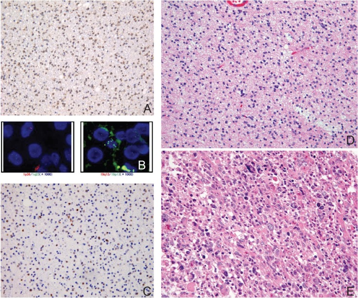
Fig. 3.
Histology of non-classic oligodendroglioma with single-positive genetic signature. A: Immunohistochemistry with isocitrate dehydrogenase (IDH)1R132H mutation specific antibody is diffusely positive in tumor cells. B: Fluorescence in situ hybridization (FISH) study showing multiple signals of both 1p36 and 19q13 indicative of polysomy. C: Immunohistochemistry with p53 shows abundant positive cells. D: Representative H&E staining section of non-classic oligoastrocytoma. The oligodendroglial component shows round but irregular nuclei. E: The recurrent tumor 2 years after the initial resection shows marked atypia and pleomorphism corresponding to anaplastic oligoastrocytoma (H&E staining).

