
Fig. 1.
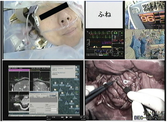
The screen of the dedicated intraoperative examination monitor for awake craniotomy (IEMAS). Upper left display: The patient’s face. Lower left display: The anatomical data obtained from the real-time updated neuronavigation system, which can be used to localize the exact position of the stimulator. Upper right display: Four different sets of data for the test object, bispectral index monitor, heart beat monitor, and general view of the operating theater. Lower right display: The view of the surgical field through the operative microscope.