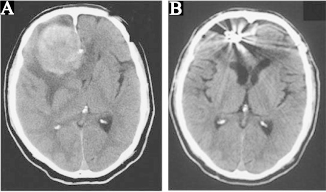
Fig. 1.

A: Preoperative computed tomography (CT) scan showing a 60-mm diameter mass in the right frontal lobe connected to the cerebral falx with partial calcification, evident cerebral edema at the periphery, and median deviation from the cerebral ventricle. B: CT scan at 1 month after surgery showing the clip artifact, disappearance of the mass and edema, and preservation of the structure of the ventricle.
