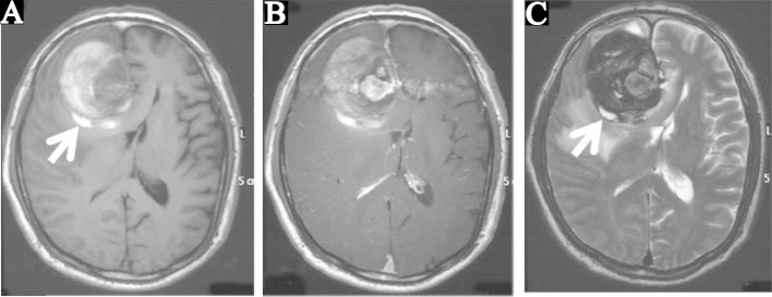
Fig. 2.
Preoperative T1-weighted (A), T1-weighted with contrast medium (B), and T2-weighted magnetic resonance images (C). Intraaneurysmal thrombus was primarily present as hyperintensity on the T1-weighted image and hypointensity on the T2-weighted image. Heterochronous thrombi in the hyperintensity were confirmed on both T1- and T2-weighted images in part of the aneurysm wall (arrows).

