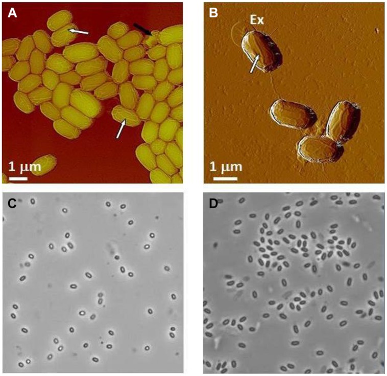
FIGURE 9.

Characterization of Bacillus spores. (A,B) AFM images of native air-dried spores. (A) Height image of B. anthracis Sterne spores and (B) amplitude image of B. thuringiensis spores. In both images surface ridges extending along the entire length of spores (several surface ridges noted by white arrows) are seen. In (A) a collapsed spore is indicated with a black arrow. In (B) an exosporia is indicated with Ex. (C,D) Phase contrast microscopy images of B. anthracis Sterne spores (C) and B. thuringiensis spores (D).
