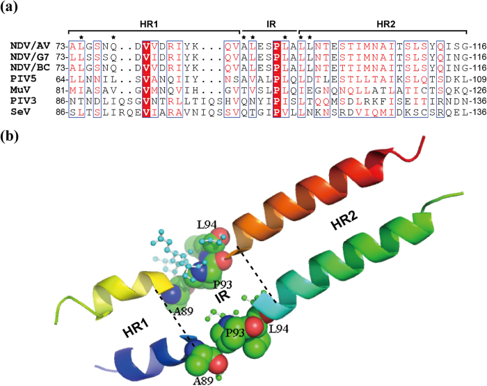
Figure 1. Helical structure of the HN stalk region.
(a) Sequence alignment of the stalk of HN proteins from three different NDV strains and four other paramyxoviruses. Conserved residues (>70%) are boxed; completely conserved residues are highlighted in red. Fusion promotion residues are indicated by black asterisks. (b) Three-dimensional structure of the NDV-AV HN stalk generated by PyMOL software (Version 1.5, Schrödinger). Residues in the IR are displayed in ball and stick form, whereas the point mutations examined in this study are showed in sphere form. The structure was derived from the crystal structure of the NDV-AV HN protein reported by Yuan et al.5.

