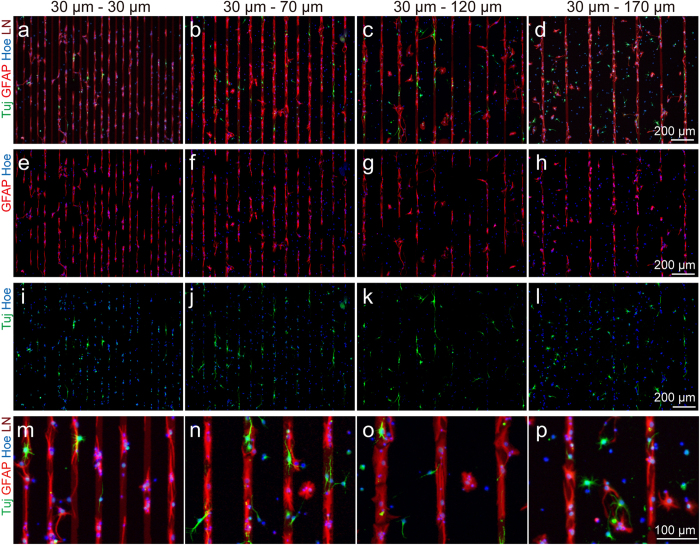
Figure 2. Differentiation and alignment of aNSCs on LN patterned culture substrate.
(a-d) Fluorescence micrographs of GFAP, Tuj-1, and nucleus stained aNSC at 6 DIV on the LN stripe patterns with 30, 70, 120, 170 μm of spacing. (e-h) Fluorescence micrographs of GFAP and nucleus stained aNSC at 6 DIV on the LN stripe patterns with 30, 70, 120, 170 μm of spacing. (i-l) Fluorescence micrographs of Tuj-1 and nucleus stained aNSC at 6 DIV on the LN stripe patterns with 30, 70, 120, 170 μm of spacing. (m-p) Fluorescence micrographs at high-magnification of GFAP, Tuj-1, and nucleus stained aNSC at 6 DIV on the LN stripe patterns with 30, 70, 120, 170 μm of spacing.

