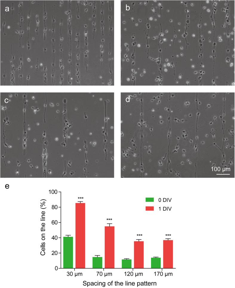
Figure 4. The migration of aNSC on the engineered culture substrate in early stage.
(a-d) Phase-contrast micrographs of aNSC on the LN stripe patterns with 30, 70, 120, 170 μm of spacing at 1 DIV. Even though LN stripes were not indicated, the stripes were identifiable owing to the preferential localization of aNSCs on LN stripes. (e) Distribution of cells on LN line pattern at 0, 1 DIV (mean ± S.E., n = 10, *p < 0.001).

