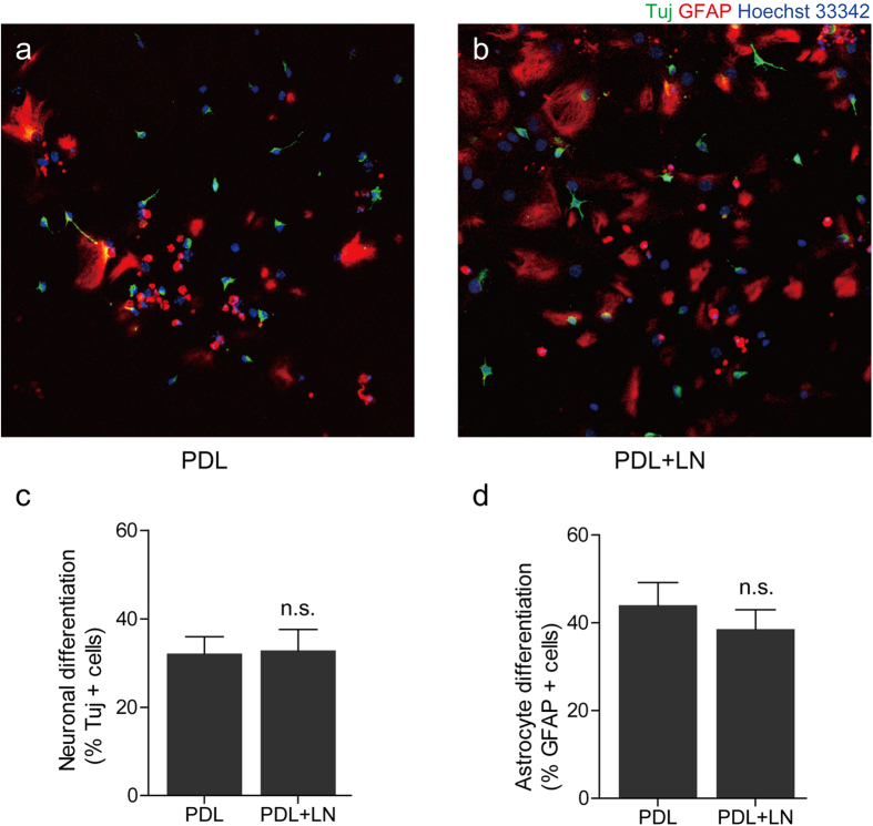
Figure 6. The differentiation of aNSC on laminin coated substrate.
(a) Fluorescence micrograph of aNSCs on PDL coated substrate at 6 DIV. (b) Fluorescence micrograph of aNSCs on PDL and LN coated substrate at 6 DIV. (c) Neuronal differentiation rate (mean ± S.E. n = 10) of aNSCs on each substrate. (d) Astrocyte differentiation rate (mean ± S.E. n = 10) of aNSCs on each substrate. n.s.: not significant.

