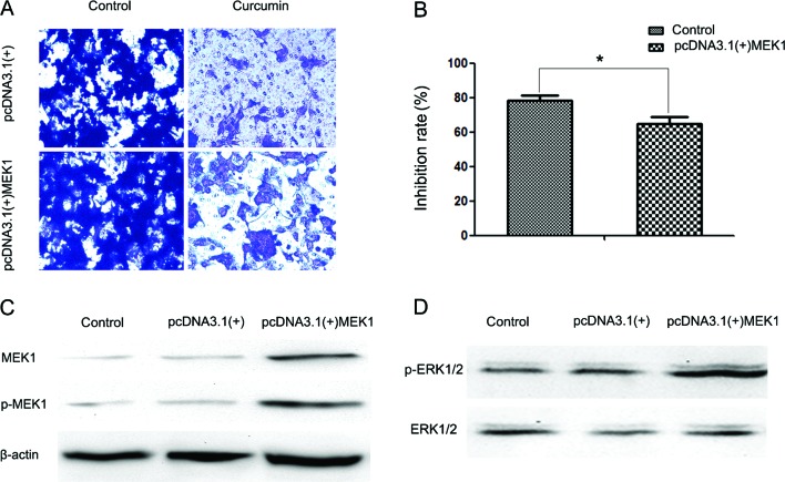
Figure 5.
Ectopic expression of MEK1 reversed the inhibitory effect of curcumin on cell invasion. (A) Transwell assays were performed to evaluate the anti-metastatic effects of curcumin (10 µM) on two groups of cells transfected with the indicated plasmids (empty vector pcDNA3.1(+) or pcDNA3.1(+)-MEK1). Magnification, x100. (B) Inhibition rates of curcumin on the two groups of cells. The inhibition rate is expressed as the inhibition cells as a percentage of the control (the control group of cells transfected with empty vector pcDNA3.1(+) or with pcDNA3.1(+)-MEK1, without treatment of curcumin). Values represent the means ± standard deviation of three independent experiments performed in triplicate. *P<0.05 and **P<0.01 compared with the control group. (C) Western blotting was performed to detect the expression of MEK1 and p-MEK1 in three groups of HEC-1B cells: no transfection (control), cells transfected with empty vector pcDNA3.1(+) (negative control group), and cells transfected with pcDNA3.1(+)-MEK1 (positive group). (D) Western blotting was performed to detect extracellular signal-regulated kinase (ERK) activity in the three groups of HEC-1B cells.

