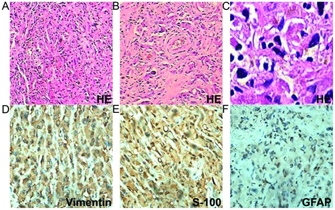
Figure 4.
Tumor pathology. (A) Sheets of tumor cells (H&E staining; magnification, x40). (B and C) Rhabdoid cells with eccentric prominent nuclei and abundant eosinophilic cytoplasm (H&E staining; magnification, x200 and x400, respectively). (D) Tumor cells demonstrating diffuse cytoplasmic immunoreactivity for vimentin (magnification, x400). (E) Tumor cells demonstrating cytoplasmic immunoreactivity for S-100 (magnification, x400). (F) Tumor cells demonstrating cytoplasmic immunoreactivity for GFAP (magnification, x400). H&E, hematoxylin and eosin; GFAP, glial fibrillary acidic protein.

