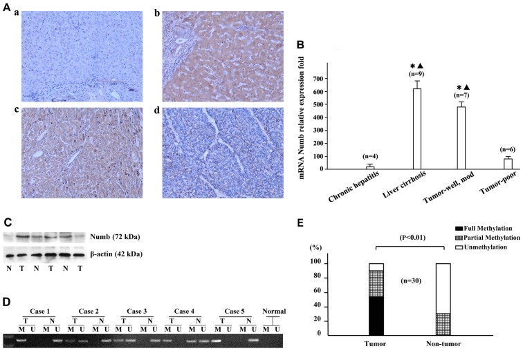
Figure 1.
Expression of Numb in samples from hepatocellular carcinomas (HCC) patients. (A) Expression of Numb in non-tumorous and tumor liver tissues detected by immunohistochemistry; (a) chronic hepatitis, (b) liver cirrhosis, (c) moderately differentiated HCC, (d) poorly differentiated HCC. Magnification, ×100. (B) Reverse transcription-quantitative polymerase chain reaction (PCR) was used to examine Numb mRNA expression from 13 paired HCC tissues and their non-tumor counterparts. The results (mean ± standard deviation) were normalised for β-actin expression. Statistical analysis was performed comparing liver cirrhosis, well- and moderately differentiated tumors, and poorly differentiated tumors with chronic hepatitis; *P<0.05; and liver cirrhosis, and well-and moderately differentiated tumors compared with poorly differentiated tumors; ▲P<0.05. (C) The representative expression of Numb as assessed by western blot analysis. Lane 1, chronic hepatitis B; lanes 2 and 4, moderately differentiated tumors; lanes 3 and 5, liver cirrhosis; lane 6, poorly differentiated tumor. (D) Methylation of Numb promoter in HCC tissues was determined by methylation-specific PCR (MSP). (E) MSP showed that the methylation of Numb was significantly detected in HCC tissues (T) compared with the corresponding non-tumorous tissues (N); n=19, P<0.01. M, methylated DNA; U, unmethylated DNA.

