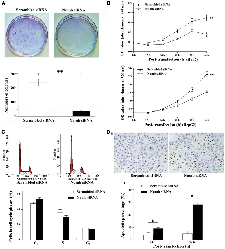
Figure 3.
Oncogenetic potential of Numb in hepatocellular carcinomas (HCC). (A) As shown by representative dishes, Numb depletion significantly inhibited the colony formation of Huh7 cells in soft agar culture. Quantitative analyses of colony numbers are shown in the lower panel. Values are the mean ± standard deviation (SD) of ≥3 independent experiments. (B) MTT assay was performed in Huh7 and HepG2 cells. Growth curves of Numb small interfering RNA (siRNA) cells were compared with the scramble cells, respectively. The results are expressed as mean ± SD of ≥3 independent experiments. (C) Flow cytometric analysis showing that Numb siRNA for 24 h increased the G0-G1 phase fraction and decreased the S phase fraction when compared to the scramble treated cultures in Huh7 cells. (D-a) Representative images of TUNEL staining. A larger quantity of apoptotic cells were detected following treatment of Huh7 cells with Numb siRNA in comparison with that of the scramble cells. (D-b) The apoptotic index was compared between Numb siRNA and scramble cells (right panel).**P<0.01, *P<0.001.

