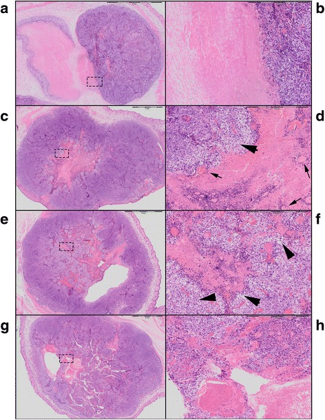
Fig. 6.

Digitalized hematoxilin and eosin stained sections of the mice tumors. 1× magnification of the tumor control (a), thermal (c), intermediate (e), and mechanical high intensity focused ultrasound treatment (g). Magnification of 6.6× of the corresponding rectangle on the left: a, b well demarcated “old” necrosis without ballooning degeneration surrounding the necrotic area. c, d Large pink necrotic area in the center of the tumor surrounded with ballooning degeneration (arrow head) and dying cells and remnants of vessels (arrow) within the necrotic lesion. e, f Fractionated tissue with islands of necrosis and ballooning degeneration (arrow heads) of cells. g, h Fractionated tissue with bleeding and formation of a cavity filled with erythrocytes
