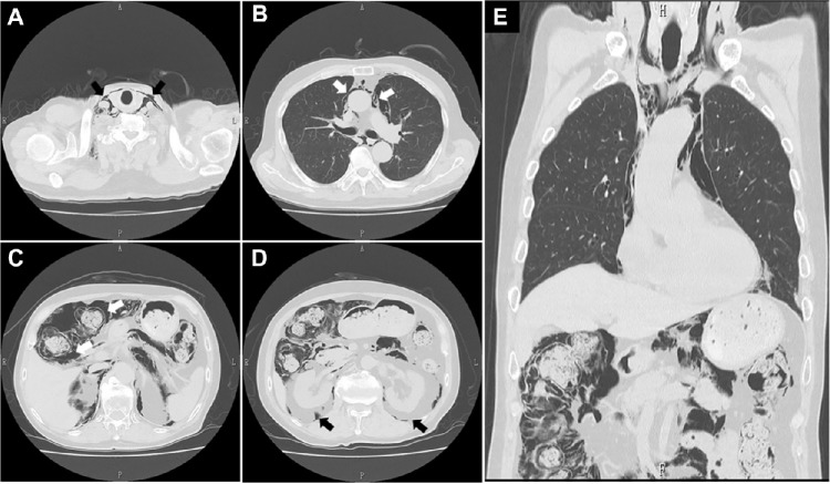
Figure 4.
CT scans of the chest and abdomen. Abnormal air accumulations characterized by radiolucent areas (black or white arrows in each panel) were observed in the cervical regions (A), mediastinum (B), intramural parts of the ascending colon (C) and mesentery, along with the retroperitoneum (D). A coronal reconstructed image (E) failed to show free air.

