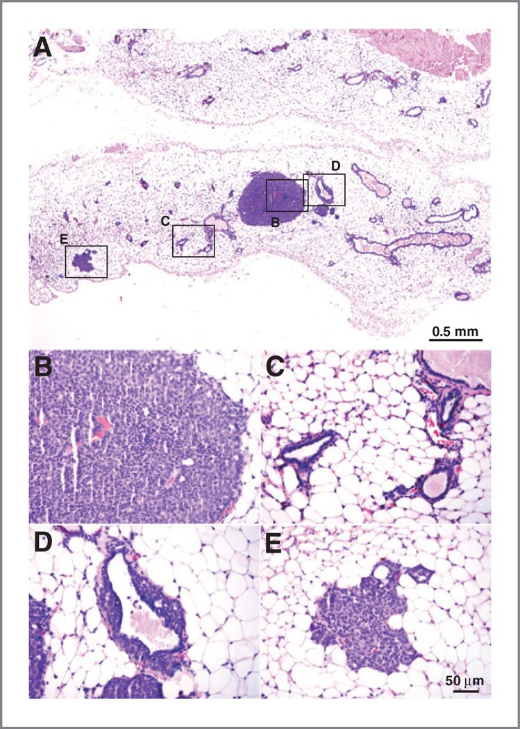
Figure 4.
Detection of microtumors in Ad-CX3CL1–treated mice. A, panoramic view of hematoxylin and eosin-stained mammary glands without macroscopic tumors from an Ad-CX3CL1–injected mouse. Selected areas are shown at higher magnification in tumor mass (B), normal glands (C), and glands with intraductal epithelial proliferation (D). E, some glands distant from the tumor showed characteristics similar to the hyperplastic foci described above.

