
Figure 2.
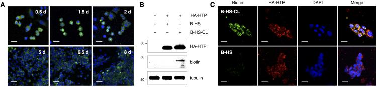
Extended cell-surface display by HTP anchoring. A) CHO cells stably expressing HTP were functionalized with a single treatment of F-CL (shown in green) and imaged over 8 days. Cell nuclei were co-stained at each time point with DAPI (shown in blue). B) Western blot detection and C) fluorescence imaging of hemagglutinin (HA)-tagged HTP and biotinylated HS. Stably transfected CHO cells were labeled with biotinylated HS with or without the chloroalkane linker (B-HS-CL or B-HS, respectively). Tubulin was used as a control for equal protein loading in B. Scale bars represent 20 μm.