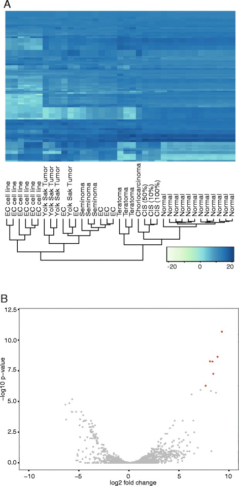
Fig. 4.

miRNA expression in normal and TGCT samples. a Heatmap showing the expression data of the 100 most highly expressed miRNAs (variance stabilization transformed data). The dendrogram (bottom) indicates clustering according to expression profiles. Heatmap colors represent relative miRNA expression as indicated in the color key (bottom right). b Volcano plot indicating the relationship between the Log2 fold change and p-values (Benjamini-Hochberg adjusted) for all miRNAs when comparing TGCT with normal samples. The CIS samples are included in the TGCT group. Red color coding indicates a significance level of p < 10−8 on a –log10 scale and log2 fold change above 5/below −5
