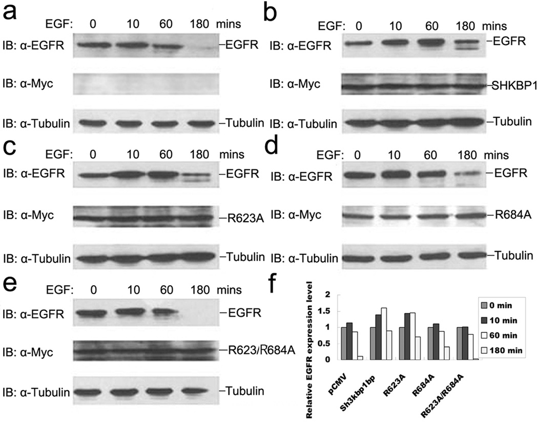
Figure 3. SHKBP1 inhibited EGFR degradation after EGF stimulation.
HeLa cells were transfected with indicated expression plasmids: pCMV (a), Myc-SHKBP1 (b), Myc-R623A (c), Myc-R684A (d) and Myc-R623A/684A (e), respectively. Thirty-six hours post-transfection, cells were starved in serum free DMEM for 12 hours. The cells were then stimulated with 100 ng/ml EGF for the indicated times. Lysates were prepared and analyzed by western-blot with anti-EGFR, anti-Myc or anti-α-tubulin antibodies. The relative level of EGFR (a–e) was quantified by densitometry scan and graphed in (f).

