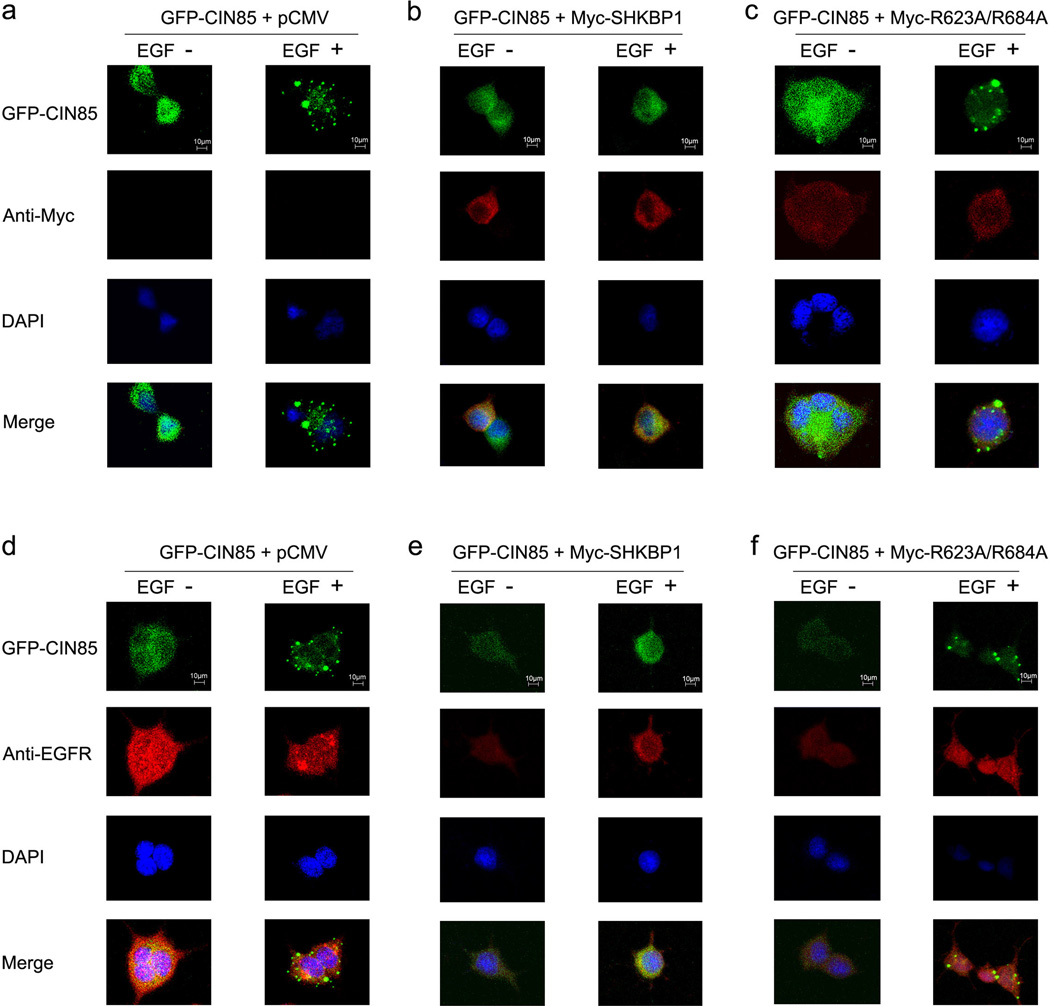
Figure 4. SHKBP1 disturbed the translocation of CIN85 to EGFR degradation vesicles.
HEK293T cells were co-transfected with GFP-CIN85 plasmid and control pCMV mock vector (a, d), Myc-SHKBP1 (b, e) or Myc-R623A/R684A (c, f). Thirty-six hours post-transfection cells were starved in serum free DMEM for 12 hours. The cells were left unstimulated or stimulated with hEGF for 10 minutes. Immunofluorscence studies on cells were performed with antibodies against Myc tag (a, b, c) or EGFR (d, e, f), and the nucleus were stained with DAPI. The merged pictures were shown in the bottom panel, and the staffs (10µm) were shown in the upper panel.

