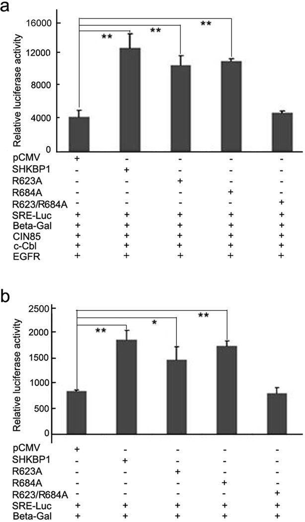
Figure 5. SHKBP1 enhanced EGF induced SRE transcription activity.
a HEK293T cells were co-transfected with EGFR (100ng), c-Cbl (100ng), CIN85 (100ng), SRE-Luc (100ng), β-gal (50ng), and SHKBP1 or its mutants plasmids (300ng). Thirty-six hours post-transfection cells were starved in serum free DMEM for 12 hours. The cells were then stimulated with EGF for another 12 hours and processed for luciferase assay.
b HeLa cells were co-transfected with SRE-Luc (200ng), β-gal (100ng), and SHKBP1 or its mutants plasmids (300ng). Thirty-six hours post-transfection cells were starved in serum free DMEM for 12 hours. The cells were then stimulated with EGF for additional 12 hours and processed for luciferase assay. The luciferase activity was measured in triplicated samples and expressed as the mean ± s.d. Student’s t-test was performed to compare the statistical difference between each group to pCMV control group; ‘**’ indicates p<0.01, ‘*’ indicates p<0.05.

