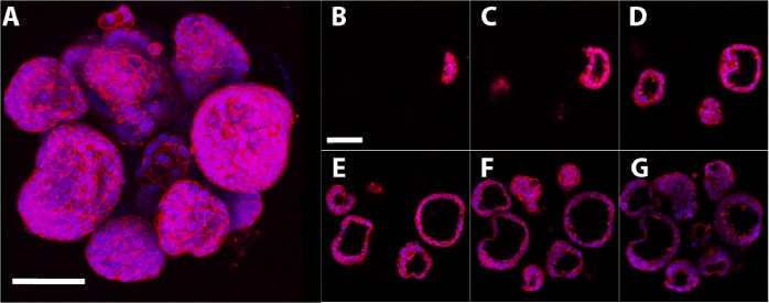
Fig 2. 3D structure of 7-day MCF-7 microtissues.
Intact MCF-7 microtissues were stained with rhodamine phalloidin (red; F-actin) and DAPI (blue; nuclear stain) and imaged on a confocal microscope, with steps taken in the Z direction every 5μm. Individual images were compiled to make a maximal projection image, demonstrating the morphology of the microtissues (A). Sequential images at intervals of 15μm in the Z direction demonstrate that microtissues contain luminal spaces (B-0μm, C-15μm, D-30μm, E-45μm, F-60μm, G-75μm). Scale bar = 80μm.

