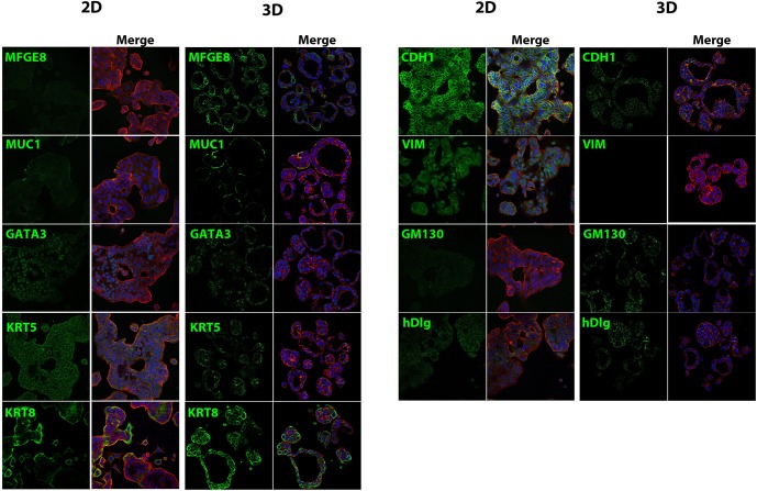
Fig 3. Expression of luminal and epithelial markers in 3D MCF-7 microtissues.
Three day monolayer cultures and cryosections of MCF-7 cells grown for 7 days in agarose hydrogels were stained for epithelial and luminal markers (green), with f-actin rhodamine phalloidin (red) and Hoechst 33342 nuclear stain (blue) counterstains. Monolayer cultures exhibited diffuse staining for milk fat globule EGF8 (MFGE8) while 3D cultures showed intense, localized staining at the basal surface. Mucin 1 (MUC1) staining was faint and localized to select cells in 2D cultures with more intense basal staining in 3D microtissues. Expression of the luminal transcription factor trans-acting T-cell-specific transcription factor (GATA-3) was localized to the nucleus in both 2D and 3D cultures. Keratin 5 (KRT5) staining was diffuse in 2D cultures and more concentrated in specific cells in 3D cultures. There was high expression of keratin 8 (KRT8) in monolayer cultures, and throughout the microtissues (S, T). MCF-7 cells cultured in 2D demonstrated diffuse staining throughout the cytoplasm for e-cadherin (CDH1), while cells grown in agarose hydrogels displayed localized at cell-cell contacts. Mesenchymal marker vimentin (VIM) was present in the cytoplasm and nucleus of monolayer cultures and not detectable in 3D cultures. The Golgi marker GM130 displayed diffuse staining in monolayer culture but was punctate in MCF-7 microtissues, with localization at the apical surface in select cells, and in other was basolaterally oriented. Human disc large (hDlg) displayed diffuse staining in 2D cultures, but was expressed at cell-cell contacts and the basal surface in 3D cultures.

