Fig. 7.

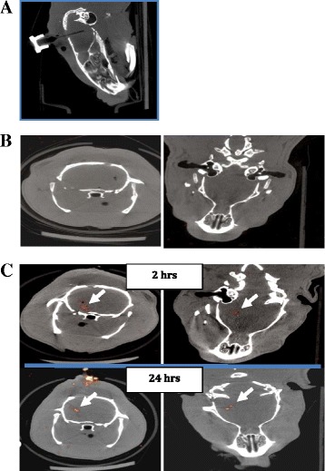
Repetitive in vivo SPECT imaging of NIS-Hipp-NSCs. a CT scan of the rat head showing the placement of the cannula used to deliver 99mTc into the brain. b SPECT imaging of the brain of a rat that had received 99mTc and no cells. c SPECT signal recorded from the rat brain 2 and 24 h after NIS-Hipp-NSC intracranial injection following delivering of 99mTc. Transverse and coronal slice orientations are shown. CT computed tomography, NIS-Hipp-NSC hippocampus-derived neural stem cell expressing the human sodium iodide symporter, SPECT single-photon emission tomography, 99m Tc technetium-99m
