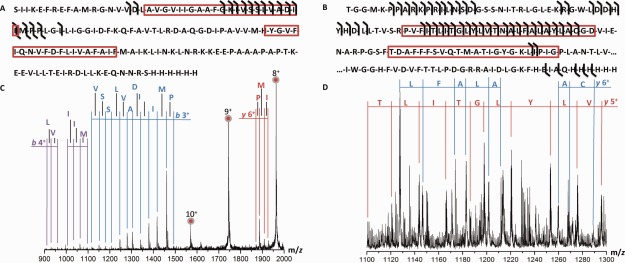
Figure 3.

Primary structure of membrane proteins investigated using top-down CID. (A) Sequence of MscL G22C with b- and y-fragments indicated and (B) Excerpt (1–112 and 265–300) from the sequence of Kirbac3.1. Predicted membrane spanning regions are indicated with a red box. (C) Part of the spectra observed for MscL and (D) Kirbac3.1 displaying CID fragments.
