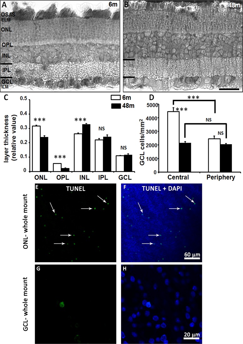
Fig 1. Light and confocal fluorescence micrographs illustrating the morphology of the retina in young and adult degus.
Retinal sections from young and adult degus were stained with Toluidine blue (A, B). Quantification of retinal layer thickness (C) and ganglion cell density in young and adult degus (D). TUNEL labeling of the adult retina showed apoptotic cells in the ONL (E, F) but not in the GCL (G, H) in the retinal whole mount. Abbreviations: 6 months old (6m), 48 months old (48m), outer and inner segment of the photoreceptors (OS/IS), external limiting membrane (ELM), outer nuclear layer (ONL), outer plexiform layer (OPL), inner nuclear layer (INL), inner plexiform layer (IPL), ganglion cell layer (GCL), inner limiting membrane (ILM). Scale bar = 20 μm.

