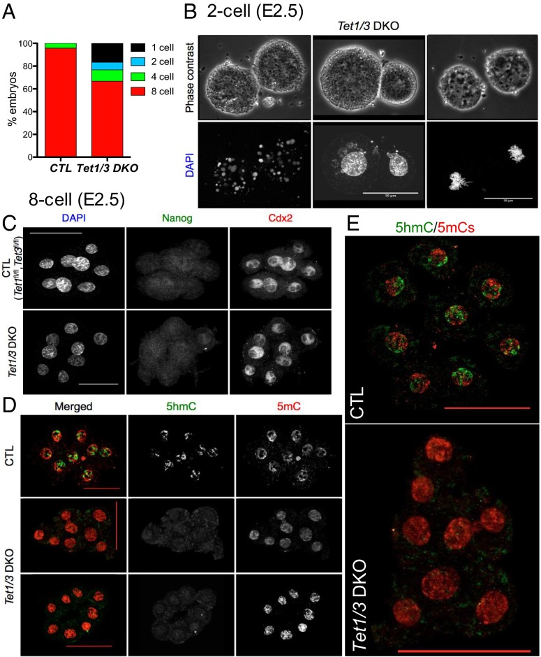
Fig. 1.
Analyses of control and Tet1/3 DKO two-cell and eight-cell embryos. (A) Bar graph showing the percentage of one-cell, two-cell, four-cell, and eight-cell embryos in control (CTL) and Tet1/3 DKO embryos harvested at E2.5. Almost all CTL embryos have reached the eight-cell stage, whereas a substantial fraction of Tet1/3 DKO embryos show aborted or delayed development. (B) Apoptosis (Left), nuclear fragmentation (Middle), and mitotic defects (Right) in representative nonviable two-cell embryos from Tet1/3 DKO mice revealed by DAPI staining. (C) Immunocytochemistry for the lineage markers Nanog and Cdx2 does not reveal any pronounced differences between CTL and Tet1/3 DKO eight-cell embryos. Cdx2 was expressed in all blastomeres, whereas Nanog expression was typically not detected in eight-cell embryos (or occasionally detected in one or a very few blastomeres). (D) Immunocytochemistry using antibodies to 5mC and 5hmC shows global loss of 5hmC and concomitant increase of 5mC in nuclei of Tet1/3 DKO eight-cell embryos compared with CTL. (Scale bar, 50 μm.) (E, Top) Merged and enlarged images of a second control embryo. (Bottom) Tet1/3 DKO embryo from D, Middle, stained with antibodies to 5mC and 5hmC.

