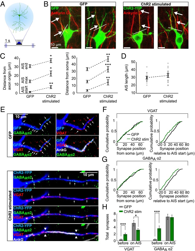
Fig. 2.
Chronic stimulation of CA1 pyramidal neurons leads to a change in AIS, but not in axo-axonic synapse position. (A) ChR2-transfected CA1 pyramidal neurons in organotypic slices were stimulated with LEDs for 48 h. (B) AnkG stain in GFP control neurons and ChR2-stimulated neurons. Arrows indicate start and end position of the AIS, white line indicates axon origin. (C) The AIS position in control and chronically stimulated neurons, measured from axon origin or from soma (n = 15 GFP cells, n = 16 ChR2-stimulated cells). (D) AIS length in control and chronically stimulated neurons (n as in C). (E) VGAT, GABAAα2, and AnkG staining in GFP control neurons (Upper) or ChR2-stimulated neurons (Lower). Arrows point to axo-axonic synapses overlapping with the AIS in 3D. The AIS position is marked with arrowheads. (F and G) Empirical cumulative distribution functions of VGAT (F) and GABAAα2 (G) puncta positions relative to the soma or to the AIS start (VGAT: n = 136 synapses, 14 cells control; n = 109 synapses, 14 cells ChR2-stimulated; GABAAα2: n = 207 synapses, 14 cells control; n = 183 synapses, 14 cells ChR2-stimulated). (H) Number of VGAT and GABAAα2 puncta observed between the soma and the AIS as well as on the AIS in control and light-stimulated neurons (n = 14 cells). All error bars represent SEM; *P < 0.05, **P < 0.01, ***P < 0.001.

