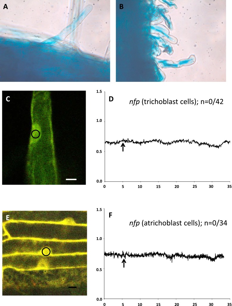
Fig. S6.
Analyses of MVA-induced ENOD11 expression and Ca2+ spiking in nfp mutants (nfp-1 and nfp-2). (A) MVA-induced ENOD11 expression in the trichoblast and atrichoblast cells of wild type. (B) MVA-induced ENOD11 expression in the trichoblast and atrichoblast cells of nfp-2. (C) Representative trichoblast cell expressing YC3.6 used for Ca2+ spiking analyses. (D) MVA failed to induce nuclear Ca2+ spiking in trichoblast cells of both nfp mutants. (E) Representative atrichoblast cells expressing YC3.6 used for Ca2+ spiking analyses. (F) MVA failed to induce Ca2+ spiking in atrichoblast cells of both nfp mutants. (Scale bar, 10 µm.) The small circle in panels C and E indicates the region of interest used for obtaining FRET and CFP channel intensities to analyze nuclear Ca2+ spiking.

