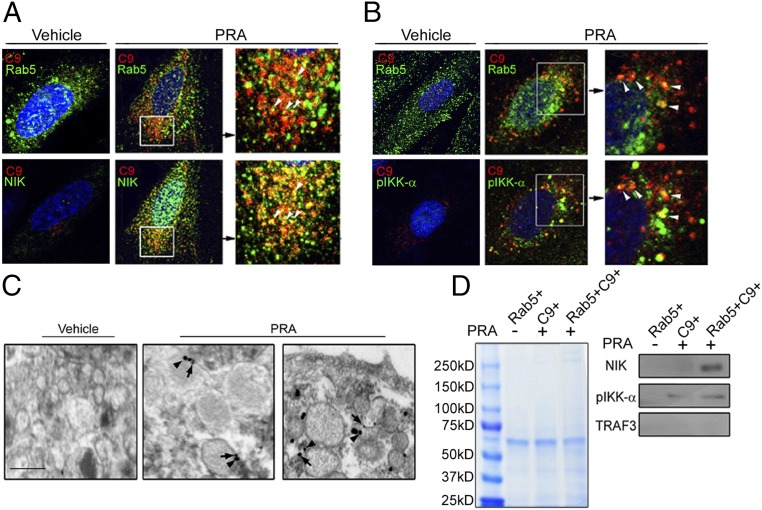
Fig. 4.
MAC+NIK+ signalosome forms on Rab5+ endosomes. (A and B) Three-color staining of NIK, C9, and Rab5 (A) and pIKK-α, C9, and Rab5 (B). (C) Immune-EM of C9 (arrowheads) and NIK (arrows) in ECs treated with PRA for 30 min. (Scale bar: 100 nm.) (D) ECs were transfected with Rab5-GFP and treated with MACs containing labeled C9-Alexa Fluor 647. Subcellular fractions containing endosomes were analyzed by Coomassie blue staining (Left), followed by Western blot analysis (Right). Each experiment was repeated two to three times, with similar results.

