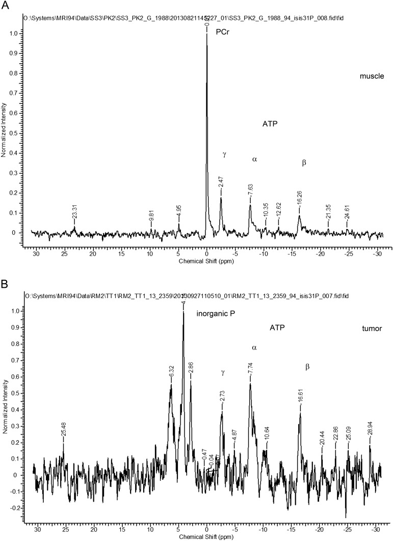
Fig. S2.
31P MRS spectra from muscle and tumor. 31P MRS spectra were acquired from BALB/neu-T mice as described in Materials and Methods and Fig. 4. (A) Spectrum from muscle (256 averages) and (B) spectrum from tumor (1,024 averages). Initially, mice were injected i.p. with 3-APP [132 mg/mL in saline, 0.2 mL; the mice indicated as “(HIGH)” received a higher dose of 333.33 mg/mL, 0.3 mL] before measurement. Because no 3-APP signal could be detected within tumor voxels (Table S1), we used inorganic phosphate; α-, β-, and γ-ATP; and PCr peaks for pH calculations.

